
The team in this service treats conditions at the back of the eye, which are treated medically using drugs, eye drops or lasers, and includes diabetic eye screening.
Conditions treated by clinicians in this service include age-related macular degeneration (AMD), retinitis pigmentosa, diabetic retinopathy, retinal blood vessel blockages and inflammation at the back of the eye (uveitis).
Let diabetes not affect your eyes
OUR APPROACH
With the increase in incidence of diabetes and increase in life expectancy, the risk of retinal disorders such as diabetic retinopathy and age related macular degeneration has gone up. We continuously make efforts to create awareness in patients suffering from diseases such as diabetes, heart conditions, high blood pressure, etc. who are more susceptible to be affected by retinal disorders. In some cases, the damage caused to the eye is irreversible. Therefore, we also conduct regular family screening clinics, diabetes screening clinics etc. so that we can identify retina disorders before they grow to a vision threatening stage.
OUR PROCESS
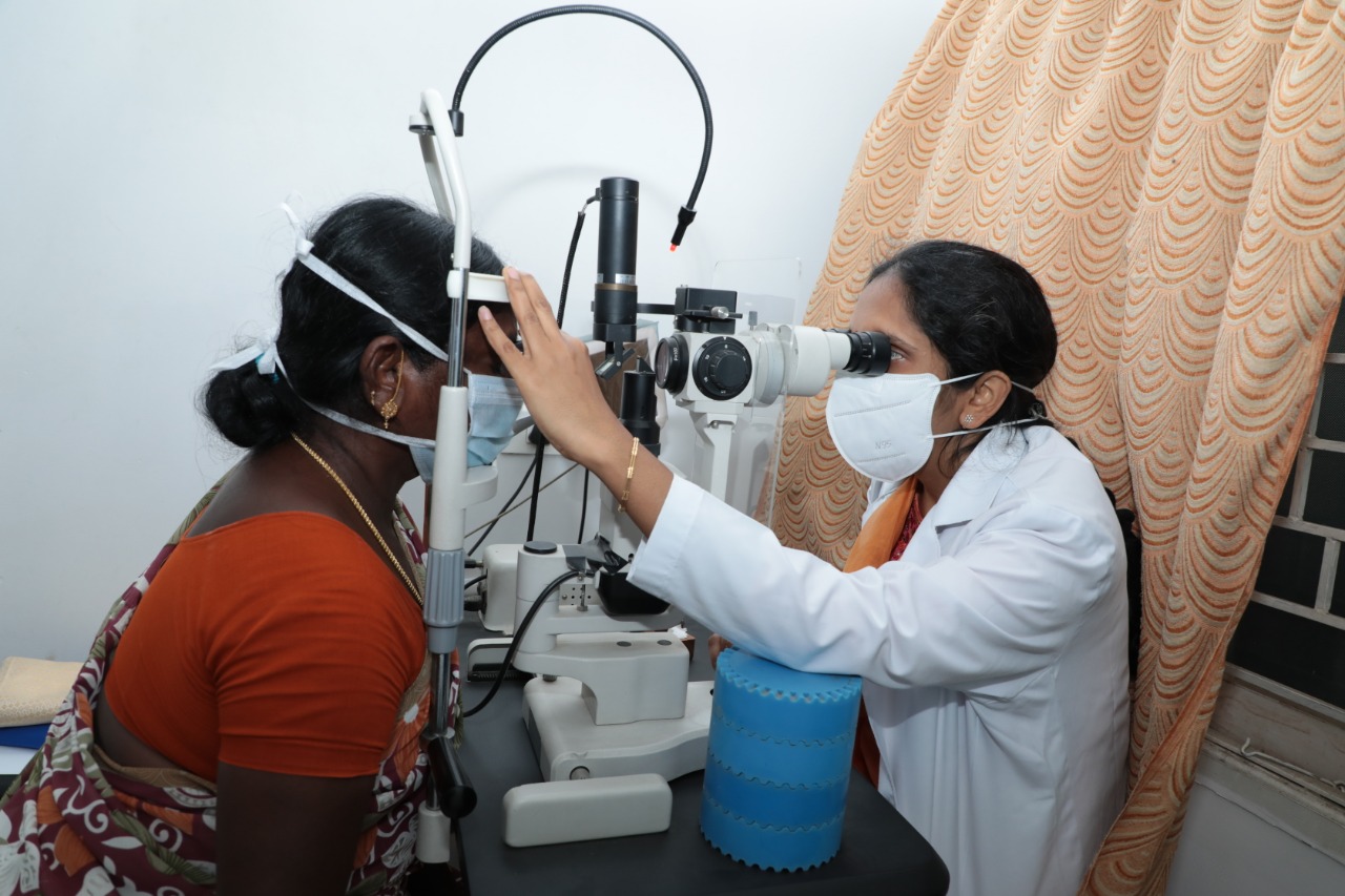
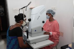
The patient’s eye is dilated and examined using advanced diagnostic technologies such as OCT, angiography, fundus photography, retinal imaging and ultra sound. A photographic documentation of the retina helps to keep a track of the improvement in the eye.
LASER PHOTOCOAGULATION
This procedure involves using laser for retinal photocoagulation. It is often used for patients with diabetic retinopathy, venous occlusions, peripheral retinal holes and lattice degenerations.
INTRAVITREAL INJECTIONS
Drugs such as Lucentis, Avastin, Ozurdex and others are injected in the eye to prevent further loss of vision after the pupil is dilated and numbed with anesthesia. These can often help in halting progression or reversing the disease pathology in diabetic retinopathy, vein occlusions or age related macular degeneration.
VITREO RETINAL SURGERY: MINIMALLY INVASIVE VITRECTOMY SURGERY (MIVS)
Vitrectomy is done to repair or prevent retinal detachment, especially when it threatens to affect the macula. We have the most advanced vitrectomy machine and the expertise to deal with complex retinal problems.
FUNDUS ANGIOGRAPHY
A small amount of yellow fluorescein dye is injected which travels to the eye, where it highlights the blood vessels. It is particularly useful in showing leaking blood vessels and highlighting where the blood supply at the back of the eye is poor. After this, photographs of the eye are taken.
RETINAL ULTRASTRUCTURE EXAMINATION (HIGH DEFINITION OCT)
This examination allows the doctor to understand the problems of patient’s retina, which might often not be identified using other optical techniques.
ULTRASOUND EXAMINATION OF RETINA
While optical techniques can reveal much about the structure of the retina, ultrasound allows imaging of the choroid and deeper tissues.
Warning Symptoms:
- Blurred, double, or distorted vision or difficulty reading.
- Floaters or spots in your vision.
- Partial or total loss of vision or a shadow or veil across your field of vision.
- Pain, pressure, or constant redness of the eye.
All diabetics are at risk of getting Diabetic Retinopathy. The risk multiplies the longer a person lives with diabetes. It has been observed that about 80% of long standing diabetics (15 years or more of diabetes) have some damage in the blood vessels of retina. Diabetic retinopathy can occur at a young age in juvenile diabetics.
Diabetic retinopathy is a silent vision stealer. In early stage
, there is hardly any symptom. Hence, a diabetic must strictly get his eye exam done every year. If detected early, vision loss can be prevented. But, once the damage is done, the effects are irreversible.
The ophthalmologist will do a comprehensive eye investigation to detect Diabetic Retinopathy. The eye exam includes visual acuity tests, eye pressure measurements and direct visualization of the retina with an ophthalmoscope. Sometimes the ophthalmologist may require more extensive imaging with technology like Fundus Fluorescein Angiography (FFA) and Optical Coherence Tomography (OCT) to capture the details of the damage caused by abnormal blood vessels & assess the severity of the condition.
Diabetic Retinopathy Treatment

Laser Photocoagulation:
It is the most common line of action for Diabetic retinopathy treatment. But remember, this can only save the existing sight level and cannot make it better. In laser treatment, the retina specialist uses laser to destroy areas of retina deprived of oxygen which helps to prevent growth of new blood vessels into these areas. It can be done in multiple sessions. In most cases, this procedure causes the new blood vessels to regress and swelling to subside. It usually takes three to four months to be fully effective.
Sometimes the new blood vessels bleed into the gel like centre (vitreous) of the eye. This condition – Vitreous hemorrhage can lead to a sudden loss of vision. If the vitreous hemorrhage is persistent then a procedure called Vitrectomy is recommended. This is a microsurgical procedure for diabetic retinopathy treatment which removes the blood and scar tissue from the centre of the eye. Many patients have improved vision after vitrectomy.
Intravitreal Injections:
These help patients with gross swelling in the macula. It may require injections into the eye of vascular endothelial growth factor (VEGF) inhibitor drug or steroid to reduce the growth of abnormal blood vessels and leakage of fluid from them. These may also be used prior to surgery to reduce the bleeding.
The choice of treatment depends on the stage of the disease, the age of the patient and the recommendations of the retina specialist.
Diabetic Retinopathy Treatment – Success Rate
Patients who have already lost vision from the disease usually do not regain the original vision. However, vision loss from complications such as bleeding into the eye or cataracts can be regained after diabetic retinopathy treatment. There is evidence that proper blood sugar control can delay and limit the progression and complications of Diabetic Retinopathy for people with diabetes.
Retinal Detachment & It’s Management
Retinal detachment is an extremely grave eye condition that happens when the retina separates from the tissue behind it. One can permanently lose vision if the retina detachment isn’t repaired promptly. Our team of expert retina specialists is well known across the country for management of even the worst case scenarios.
Retinal tears are although different than retinal detachment, but they are often the first stage leading to retinal detachment. If fluid from within the eye passes through a retinal tear that can separate the retina from its underlying tissue requiring prompt sealing of the same with lasers.
Preserve your sight for the best years of your life
Age-related macular degeneration (ARMD or AMD) is a disease that causes progressive deterioration or breakdown of the eye’s macula. The macula is that part of the retina which is responsible for your central vision, allowing you to see fine details clearly. Macular degeneration is a part of body’s natural ageing process. It is quite common after the age of 65 years.
Treatment may slow down or prevent progressive loss of vision but may not bring the lost vision back. ARMD treatment plan includes Laser therapy & PDT, Anti–VEGF injections, Combination Therapy and Low Vision Aids. At our centre we house the best infrastructure and state-of-the-art medical equipment with a pool of experienced Retina specialists so that you can be assured that you are in the best hands.
Diabetic macular oedema
What is diabetic macular oedema?
Diabetic eye disease is a leading cause of blindness registration among working age adults in India. It is caused by changes to the tiny blood vessels of the retina (the light sensitive layer at the back of the eye). In diabetic macular oedema, blood vessels leak fluid into the retina.
How does diabetic macular oedema cause vision loss?
Vision loss occurs when the fluid reaches the macula (the centre of the retina that provides sharp vision) and builds up, causing swelling. At first, you may not notice changes to your vision. Over time, diabetic macular oedema can cause your central vision to become blurred. A healthy macula is essential for good vision.
Who is at risk of diabetic macular oedema?
All people with type 1 and type 2 diabetes are at risk of diabetic macular oedema. You are at greater risk if you:
- Have had diabetes for a long time–about one in three people living with diabetes for 20 years or more will develop diabetic macular oedema
- Have poorly controlled blood sugars
- Havehigh blood pressure
- Have high cholesterol levels
- Smoking
- Pregnancy
Large studies have shown that people who have well-controlled blood sugar, blood pressure and cholesterol levels, and do not smoke are less likely to develop diabetic macular oedema.
How to reduce the risks of diabetic macula oedema?
To reduce the risk of diabetic macular oedema, it is important not to smoke and to ensure that your blood sugar, blood pressure, and cholesterol levels are well controlled.
How is diabetic macular oedema detected?
Diabetic macula oedema may be detected during your annual eye screening visits, which are offered to all patients with diabetes. Digital photographs of your retina may show signs of early diabetic macular oedema. You may not notice any changes in your vision at this stage. If diabetic macular oedema is detected, you will be referred to the retina clinic.
What happens when I attend the medical retina clinic?
You will have a comprehensive eye examination that includes:
- Visual acuity test:A sight test that measures how well you see at different distances
- Eye pressure test:A test that measures the pressure of your eyes –numbing drops may be used as part of this test
- Dilated eye examination:Drops are placed in your eyes to dilate (widen) your pupils so that the back of your eyes can be examined You may also undergo tests such as a:
- Fluorescein angiography, a diagnostic test which involves the injection of fluorescein (yellow) dye into your bloodstream via a vein in your hand or arm, followed by a series of retinal photographs taken over several minutes. The test gives your doctor more information about the condition of your retina and this helps decide which treatment is most appropriate.
- Optical coherence tomography (OCT) measures the amount of retinal swelling (macular oedema) which, like fluorescein angiography, helps decide which treatment is most appropriate. OCT is also used to monitor your retina over time and to show how effective treatment may have been. It is effectively ‘optical ultrasound’, a non-invasive test, using reflections from within your retina to provide a cross-sectional picture of the retina.
Anti- VEGF intravitreal injection treatment
This is written for patients who might have treatment with anti-angiogenic (anti-VEGF) drugs via an injection into the eye. This treats certain retinal conditions which cause abnormal blood vessels to grow and leak under the retina.
Patients with these retinal conditions can lose central vision when abnormal blood vessels bleed or leak fluid under the retina at the back of the eye. A series of injections of anti-VEGF medicines are given into the back of the eye to stop these blood vessels growing and help control the leakage. This treatment is highly effective in preserving central vision in many people.
In which common conditions might this occur?
- Wet age-related macular degeneration.
- Myopic choroidal neovascularization.
- Diabetic macular oedema.
- Retinal vein occlusion.
- Any other retinal condition causing fluid to leak under the retina.
What is age-related macular degeneration (AMD)?
Age-related macular degeneration (AMD) is the leading cause of vision loss in people aged 50 years or older. It involves damage to the part of the eye called the macula. The macula is a small, but extremely important area located at the centre of the retina, the light-sensing tissue that lines the back of the eye.
Sub-retinal bleeding at the macula in wetmacular degenerationThe macula is responsible for seeing fine details clearly. A person with AMD loses the ability to see fine details, both close-up and at a distance. This affects only the central vision, with the side, or peripheral, vision usually remaining normal. For example, when people with AMD look at a clock, they can see the clock’s outline but cannot tell what time it is. In a similar way, those with AMD will also lose the ability to recognise people.
What is ‘wet’ AMD?
There are two types of AMD. Most people (about 75%) have a form called ‘early’ or ‘dry’ AMD, which develops when there is a waste build-up under the macula. This is usually seen in those with normal or partially reduced vision. A minority of patients with early AMD can progress to the vision-threatening forms of AMD called late AMD. The most common form of late AMD is ‘exudative’ or ‘wet’ AMD. Wet AMD occurs when abnormal blood vessels grow underneath the retina.These unhealthy vessels leak blood and fluid, which can prevent the retina from working properly. Severe damage leads to severe permanent loss of central vision, but the eye is not usually at risk of losing all vision (going ‘blind’) as the ability to see in the periphery (side) remains. There is a less common form of late AMD called geographic atrophy, where vision is lost through the macular tissue becoming completely worn out, with no leaking blood vessels. Unfortunately, anti-angiogenic medicines cannot help this form of late AMD.
What is myopic choroidal neovascularization?
This condition occurs in people who are highly myopic (short-sighted). When someone is highly short-sighted, the retina at the back of the eye is stretched due to the increased size of the eye. This stretching can make the retina thinner and prone to splitting. When this occurs, blood vessels from the choroid (the layer of the eye behind the retina) can grow underneath the retina. These new vessels (neovascularisation) leak blood and fluid, which can prevent the retina from working properly. Severe damage leads to severe permanent loss of centralvision.
What is diabetic macular oedema (DMO)?
Diabetic macular oedema is an eye condition occurring in people with both type 1 and type 2 diabetes. Macular oedema is swelling and thickening of the macula. The macula is a small area in the centre of the retina which contains a rich collection of nerve cells sensitive to light, fine detail, and colour DMO occurs as a result of changes in retinal blood vessels in people with diabetes. Diabetes is characterised by increased levels of sugar (glucose) in the blood stream. Consistently high blood sugar can damage blood vessels, with the first signs appearing in the smallest vessels, called capillaries. The damaged blood vessels will leak, causing the build-up of excess fluid (oedema) and blood in the macula. It can lead to severe impairment of central vision in the affected eye.
What is retinal vein occlusion (RVO)?
RVO occurs when one of the retinal veins is blocked. The retina is thelight sensitive tissue that lines the back of the eye and is responsible for eyesight. RVO usually occurs when:A retinal vein is ‘pinched off’ through the pressure of an artery lying on top of the vein; or is clogged with a blood clot or atherosclerotic plaque (fatty deposit in the wall of the artery); or is blocked by some inflammatory conditions. The block can occur in the main retinal vein-central retinal vein occlusion (CRVO), or in one of the branches of the main vein-branch retinal vein occlusion (BRVO). Macular oedema is swelling and thickening of the macula. The macula is a small area in the centre of the retina that contains a rich collection of nerve cells sensitive to light, fine detail, and colour. The vein block causes the blood pressure to increase in the small retinal blood vessels which causes them to bleed and leak fluid into the retina, forming a macular oedema. The retina may also be affected by poor blood flow and inflammation. All these processes lead to decrease/loss of vision in the affected eye.
Can I have treatment with anti-angiogenic (anti-VEGF) drugs if I am pregnant or breast-feeding?
If you are pregnant or planning to become pregnant, please discuss this with your doctor before your intravitreal injection treatment. Anti-VEGF medicines should be used with caution during pregnancy. Women of child-bearing potential should use effective contraception during their treatment and for at least three months after the last intravitreal injection. If you do become pregnant whilst undergoing intravitreal injections, please inform your doctor immediately. Anti-VEGF medicines are not recommended during breast-feeding because it is not known whether the medicine passes into human milk. Ask your doctor for advice before treatment.
Who else should not be treated with anti-VEGF?
You should not be given anti-VEGF treatment if you have any of the following:
- allergy to anti-VEGF or any of its ingredients.
- an infection in or around either eye or severeinfectionanywhere in your body.Anti-VEGF should be used with caution in patients who have had a heart attack or stroke in the last three months, or who have uncontrolled angina or uncontrolled high blood pressure. Please ensure you inform the doctor if there are any changes in your medical condition.
How does an anti-VEGF injection prevent sight loss?
Anti-VEGF medicines stop the abnormal blood vessels growing, then leaking and bleeding under the retina. This prevents or limits damage to the retinal light receptors and loss of central vision. These medicines are effective in preventing further central vision loss in up to 90% of treated eyes.
Is anti-VEGF treatment right for me?
Your ophthalmologist will advise if the treatment is appropriate for you and which anti-VEGF medicine will be used. Only patients with active leaking of blood and fluid can benefit from it. The treatment that’s right for you will depend on the specific condition of your central retina (macula), your vision and whether there is scarring at the macula. We perform scans and photographs of the eye which show us the different layers of the retina. These scans can show us if there is blood or fluid present within the retinal layers and help us decide on your treatment.
How is the treatment given?
The drug is injected into your eye with a fine needle. Minimal discomfort is to be expected (equivalent to having blood taken from your arm). The procedure takes five to seven minutes, but the injection itself is over in less than 20 seconds. The injection is given with you lying down comfortably on the couch inside the operation theatre. Firstly, local anaesthetic drops are applied to numb your eye and minimise discomfort. Then, your eyelids and surface of the eye are cleaned to prevent infection. Your face and the area around your eye will be covered by a small surgical sheet (a drape) to keep the area sterile. A small clip (speculum) will be used to keep the eye open (see picture below). The injection site is marked with callipers and your eye is stabilised with forceps or a cotton bud. A few seconds later, the injection is given. The injecting clinician will use lubricating drops after your injection. Your vision is assessed post injection by checking you can see hand movements or can count fingers. The image on the next page shows an eye draped with a speculum in place. The cotton bud is stabilising the eye while the injection is being given.
Who will give the injection?
These injections will be given either by an ophthalmologist.
What are the side effects of injections?
As with any medical procedure, there is a small risk of complications following anti-VEGF treatment.
Some common side effects that could occur include:·Red eye (there is usually a bleed or bruise on the white part of the eye at the site of injection, which clears in a week or two).·Sore and gritty eye (slight ache and discomfort lasting a day or two).·‘Blobs’ or ‘small specks’ in your vision (‘floaters’) might be seen for a few days after the injection. You may also experience transient flashing lights or swirls of light immediately after the injection. It is important to note that most of the discomfort relating to injections is due to the use of Povidone Iodine antiseptic. This is a vital part of the injection process and reduces the risk of infection inside the eye.
There are no special precautions following intravitreal injections and you will be able to travel, but please avoid getting water into your eye or swimming for the first few days afterwards.